December 2025
Settings Tip: Frame Rate
Are you using your ultrasound for more than just bovine repro—maybe scanning a dog or an equine neck?
If so, you may want to take a moment to adjust your settings. Small changes can make a big difference in image quality, and one important setting to look at is frame rate.
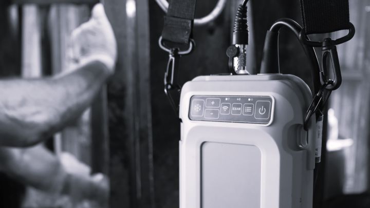
Frame rate in ultrasound refers to how many images (frames) the system captures and displays each second. Increasing the frame rate can produce smoother, more real-time motion, which is especially helpful for applications outside of standard repro work. Most of our machines default to 1 frame per second to eliminate any lag on the extension arm, but for other uses, increasing the frame rate can result in a smoother-looking image.

Merry Christmas and Happy New Year!
I love this time of year—from Thanksgiving, which gently ushers in the holiday season with a reminder of all we have to be grateful for, to the close of the year when we reflect on where we’ve been and look ahead to what’s next. Writing this letter is a sweet tradition for me, and I truly look forward to it each year. (Now, does anyone actually read it or care? I’m not entirely sure 😉.)
2025 at ReproScan has been a year of good change. We’ve had some staff changes, and they’ve been incredibly positive. You may not always see it, but behind every ReproScan product is a team of people who genuinely care about the animal health industry and about supporting you to the very best of their ability.
At the same time, 2025 has felt a bit like a rollercoaster. Between international markets, shifting policies, and the beef industry itself, I think we all ended the year with a little whiplash from the twists, turns, and ups and downs.
One bright spot in the office has been the addition of a new office puppy, Trigger. He brings plenty of laughter—especially when we’re all figuratively chasing our tails during busy season and he decides to join in literally.
Training courses continue to be a highlight for all of us. We truly enjoy spending time with you and hope that our days together help set you up for success with your preg-checking work.
Overall, it has been another good year, and we are grateful for the role you play in that. Whether we meet you at a tradeshow, talk with you over the phone, or follow up after a sale, we genuinely enjoy the connection. Thank you for helping make 2025 another blessed year for the ReproScan family.
Cheers (or as my almost two-year-old daughter says, “cheewrs”), and here’s a fond farewell to 2025 and a warm welcome to 2026. I pray that amidst the hustle and bustle of Christmas you will remember the true meaning of this holiday, that God sent his son, Jesus, to die for our sins, offering us hope eternal.
“Therefore, preparing your minds for action, and being sober-minded, set your hope fully on the grace that will be brought to you at the revelation of Jesus Christ.” -1 Peter 1:13 ESV
Merry Christmas!
–Elle
November 2025

Doc Talk with Dr. Terhaar
Doc Talk with Dr. Terhaar
Watch the video below as Dr. Terhaar explains what can be seen on the ReproScan 2.0 Monitor!
As explained, Dr. Terhaar is taking a moment to study a fetus that is laying in a unique position. When a fetus is laying in a certain way, or their body is showing certain signs, it can mean something is specific. For this fetus, Dr. Terhaar took a couple extra seconds to ensure it was not schistosomus reflexus.
An ultrasound gives you peace of mind in situations as such to determine if the fetus is healthy or other intervention is needed.
Here are some of the images you may see while preg-checking, which in turn can give you instant data to make a decision for your herd**
This ultrasound capture detected multiple fetuses. This allows a decision to be made such as where this cow calves. A rancher may choose to calve in the calving pen instead of a pasture.
This ultrasound captures shows a dead fetus in the womb. This information is vital to not only herd health, but also for the rancher/farmer as a cow may be “recycled” through breeding, so a whole breeding season isn’t lost.
Evidence of granulosa tumor cells. A granulosa cell tumor is a benign tumor of the ovary that arises from granulosa and theca cells — the same cells that are responsible for hormone production.
Two fetuses can be seen at 30 days. As stated above, finding multiple fetuses is very important. A young heifer may need more intervention, whereas an larger frame cow may be able to calve on her own.
This video confirms this fetus is a bull calf. The determination of fetal sex can be a value-adding tool to any operation. For those who plan sales, an ultrasound gives them more data to use to plan. If a cow consistently produces high-value heifer calves, you can prioritize her or her embryos for replacement heifers.
This video also shows the ear of the calf moving, demonstrating just how detailed ultrasound images can be
Ultrasounds can help you detect many things. In this case, an infection was found. Infections can be caused by an array of things and can affect many things as well. Your cows may not cycle, they could develop inflammation such an endometritis, embryo survivability could diminish, and much more.
Checking Long-Bred, Deep-Bodied Cows with the ReproArm: A Practical Guide
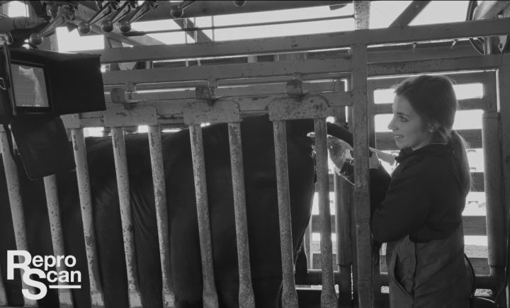
It’s that time of year…the weather is getting cooler, and the cattle are longer bred. These can be much more challenging. Late-gestation cows are a different game compared to 45–90-day pregnancies. As pregnancy advances, the uterus drops ventrally and cranially (forward and down), fluid increases, and anatomy stretches away from easy reach. Add a deep bodied and fat animal, and you’re scanning farther, through more tissue, at steeper angles. The good news: with a ReproArm and a few smart setting tweaks, you can do it.
What “Success” Looks Like Late Gestation
- Confirm pregnancy beyond doubt- at this stage you get to see cotyledons which makes it quicker.
- Good Rule: “Never call a cow open unless you visualize open uterus.” This will ensure that you do not call a cow open based on the absence of a pregnancy. That is when longer bred animals can be missed.
- If you do not see open uterus, don’t guess. Your safety double check is to palpate.
Ensure you check down deep.
- If you are working with a group of animals that you know are longer bred, I would recommend using a preset exam that is deeper or zooming out to 18-22cm deep.
- Exam D on most of our machines works well.
- Upon entry, do not lift your wrist up dramatically while pushing. This could potentially lead to rectal perforation if you lift up with your wrist and push hard at the same time.
- Advance the probe in and then lift up. For these deep bodied, long bred animals, you may be almost to the end of the regular length ReproArm.
- As you lift up and pull back ensure you sweep left to right. Envision a clock - you are scanning from 11 to 1.
- Use broad, steady rotation rather than short quick movements.
What To Look For (Fast Targets):
- Placentomes: paired caruncle–cotyledon units; often known as buttons, these grape-like structures confirm pregnancy even if the fetus is not visible.
- Fetal parts: remember you will not be able to see the whole baby in view at this stage of pregnancy.
- Heartbeat or movement: the heart may not be visible at this point unless the fetus is laying just right.
- Fluid character: flocculation will begin to be more apparent in later stages of pregnancy. It will look like swirling debris and be a bit more echogenic.
Settings That Make the Difference (Cheat Sheet)
These adjustments will help a lot.
Depth
- Set deeper first, then refine.
Typical late-gestation working 18–24 cm. Don’t be shy about maxing out depth to find your window; then zoom in if needed.
When's the last time you gave your ultrasound a little "TLC"?
Avoid a Horror Story in the Chute and Scare Off Costly Repairs with a Little Maintenance Magic: No Tricks, Just Tips: Preventative Maintenance That Pays Off
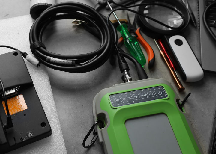
ReproScan equipment is built for durability and performance — designed to work as hard as you do. But like any hardworking tool, your ultrasound system needs periodic maintenance to stay reliable and efficient. Regular checks can prevent small issues from becoming costly repairs or unexpected downtime. Here’s what we recommend:
Every Use – Take these steps each time you use your equipment
Ultrasound & Viewing Device(s)
- Rinse probe to remove manure -> Ensure you don’t wash the machine.
- Wipe off excessive debris with paper towels.
- Clean & sanitize with a cleaning solution (Clorox Wipes work great). Do not submerge your equipment in water or use overly wet clothes.
- Wipe all equipment down with an Armor All wipe. This keeps your probe cable flexible, and your equipment looking like new!
- Put the Probe Cover on the Probe Head!
- Fully charge all equipment before storing.
Every 6 Months – Perform these quick checks
Charger & Power Supply
- Use only the original charger provided with your equipment.
- Inspect for cuts, frays, or exposed wires.
- Confirm the green light appears when plugged in.
Ultrasound Unit
- Do lights appear and blink when charging? -> They should be blinking until fully charged.
- Is the power input loose or wiggly?
- Are there cracks or missing port covers?
- Is the battery life declining?
- Any rattling sounds or image flicker (“lightning”)?
Probe
- Has the cord been pulled during use?
- Check for cuts, kinks, or exposed wires.
- Inspect the probe head and the gray surface for cracks or damage.
Monitor or Goggles
- Examine cables for damage.
- Test buttons or touchscreen responsiveness.
- Look for cracks, missing rubber covers, or a worn-out monitor bag.
- Check to ensure the monitor is still holding a charge. If you have any problems during your checks, contact our support team. We can help determine whether your unit should be sent in for repair.
Every 2 Years – Send in your equipment for a full-service check
We recommend sending in your ReproScan equipment every two years for a professional inspection. This helps us catch potential issues before they affect your work.
Our full-service check includes:

A Letter from the Office
News from the ReproScan office - Including a Small Ruminant (DVM Only) Training Course
There’s a new face around the ReproScan office—four paws, a wagging tail, and a heart full of energy! We’re excited to introduce Trigger, our new red retriever puppy.
Small Ruminant CE Course Opportunity (DVM ONLY) - Benchmark Animal Hospital
Benchmark Animal Hospital is hosting a 20 Race CE credit reproduction course. See below for all the details.
July 2025
When to Use the Shorty ReproArm vs. the Regular ReproArm
When to Use the Shorty ReproArm vs. the Regular ReproArm by ReproScan
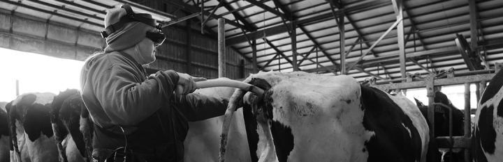
Reproductive ultrasound is all about efficiency, accuracy, and operator comfort — and having the right tools for the job makes all the difference. That’s why ReproScan offers multiple ReproArm options, including the standard Regular ReproArm and the more compact Shorty ReproArm.
While both arms are engineered with ReproScan’s commitment to durability, simplicity, and affordability in mind, each is optimized for different working conditions and cattle types. Here’s how to decide which arm is best for your scanning scenario.
Battery Best Practices:
Your ReproScan ultrasound system is built to be simple, durable, and affordable — and that includes the powerful lithium-ion battery that keeps you scanning all day. To keep your battery healthy and your scanner working at peak performance, follow these quick battery care tips:
✅Charge Smart
Charge fully, then unplug. Once your battery hits 100%, unplug it from the wall. Leaving it plugged in for extended periods can create heat buildup and gradually shorten battery life. After a day of use, plug it in to charge overnight, ensuring that the light indicates that your machine is charging. Unplug the next morning when fully charged. If you are using the 2.0 monitor, turn off the “hard shut off” switch on the back of the monitor to ensure that the touchscreen does not have a slow drain on your monitor battery.
Why to Ultrasound Over Blood Test for Preg Checking
When it’s time to preg check your cows, the tools you choose matter — not just for knowing who’s bred, but for making better management decisions across the board.
Blood testing might seem easy, but here’s the truth: You’re waiting for the results (minutes to days)… and those results can still mislead you. Studies show that residual pregnancy-associated glycoproteins (PAGs) can trigger false positives weeks or even months after a cow has lost a pregnancy.
May 2025
Don't Miss May Markdown This Year!
Warmer days are here, and so are the savings! For a limited time, take 10% off new ultrasound packages during our May Markdown Sale, happening May 12-23, 2025!
With prices on the rise, we’re giving you a rare opportunity to save on equipment that can make a long-term difference. Whether you’re looking to upgrade, add a backup unit, or start your ultrasound journey, this is the perfect time to invest.